Kobee, the Retriever-Lab mix.
Rudy, the Yorkshire Terrier.
Bailey, the Retriever mix.
Jett was also receiving the same
treatment as she had been yesterday, and she was in the closed cage receiving saline
nebulizer treatment when I arrived. She
was walking around and acting, for the most part, fine. Around mid-morning, Jen took her out to go
for a walk, and she collapsed and aspirated in the hallway. Jen rushed her back to the main area, and she,
Pat, Nancy, and Dr. Todd hooked her up to breathing machines and monitors, I
helped grab materials and do whatever they needed me to do or get whatever they
needed me to get, and Jen and Pat began giving her CPR; Jen gave her chest
compressions while Pat used an oxygen machine to pump oxygen into Jett’s lungs (at
appropriate intervals relevant to Jen’s chest compressions). After a minute or two, they were able to get
her heartbeat back, but it was too late; her brain was already gone so it didn’t
make any difference. She had a lot of
bloody fluid coming out of her mouth (the fluid that was in her lungs), so Dr.
Todd took a sample of it and was going to look at it later to see what kind
of infection it was. It was really sad,
though. That was the first time I’ve
seen a true emergency (trying to bring an animal back to life, I mean), and
since she seemed to be doing fine before it was a shocking and unexpected
happening. It was a difficult thing to
watch/help with because everyone was doing everything they could to bring her
back, but when it came down to it there was nothing that could be done. I felt completely helpless…and that was a
hard thing to experience. I realize not
every animal can be saved, though, and I’d rather have this happen than her be
in a lot of pain.
Everyone frantically working with/around Jett to bring her back to life (this was taken after I had done all I was able to do).
Other patients we had come
through today were a Poodle named Missy, a Domestic Long-Haired cat named
Lycan, and a bulldog named Buddha. Missy was getting a recheck on her left eye,
which was healing from a corneal ulcer.
Dr. Karen let me look at it through the ophthalmoscope after she did, and I could see
all the blood vessels on her cornea; Dr. Karen told me all the blood vessels
meant the ulcer was healing (as I said on day 14, the doctors would numb the
eye that had a corneal ulcer and then gently scrape it with a needle to draw
blood vessels to the ulcer, which would make it heal). Lycan had rashes on his body from most likely
an allergy (Dr. Roberta said it was most likely a food allergy). She took some hair samples from the areas of
the rash and placed them in a fungal culture to see if he had ringworm, and she
also took him into a dark room and looked at the rashes with a UV light to see
if the rashes were caused by ringworm (some forms of ringworm cause the hair in
the rashes to glow). The fungal culture
and light test both came back negative, so it most likely was an allergy. She sent Lycan home with some antibiotics,
and told the owners to be careful with what they feed Lycan (like Dr. Todd did
with Booshka’s owners on day 9). Buddha originally
came in to receive vaccines, but when Dr. Karen was doing a quick check-over on
him she discovered that he had lymph node cancer; his lymph nodes were
huge! Dr. Karen said that a lymph node
is very tiny (the size of pea, give or take a bit smaller or larger [depending
on the size of the dog]). They can’t be
seen or felt, but Buddha’s lymph nodes were at least the size of golf
balls! His owner had no idea about it,
and he was really torn up when he found out about it. Because of this, Dr. Karen didn’t give him
vaccines and instead talked to his owner about treatments and such. When she finished her appointment with
Buddha, she took me to her desk and showed me a book about lymph node cancer,
which included a normal lymph node (mostly small lymphocytes) and the different
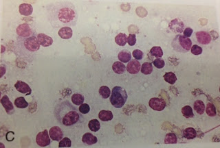
kinds of lymph node cancer that an animal can have, such as Eosinophilic Lymphadenitis
(small lymphocytes with medium lymphocytes and eosinophils [white blood cells
containing granules]), Pyogranulomatous Lymphadenitis (inflammatory cell with degenerate
neutrophils [neutrophilic white blood cells] and macrophages [large
bacteria-absorbing cells]), and having a reactive lymph node (abundance of
lymphocytes, both small and medium-sized, with plasma cells). Also, one of the night techs found a baby bird (don't know what breed) sometime yesterday, so they brought it in and were trying to keep it warm and fed. It was in a small box with a rubber glove filled with warm water, a blanket of tissues on top of the glove, the bird on the tissues, and a tissue folded as a blanket on the bird. Tracey took it home when she went home to try and see if she could raise it and keep it from dying, but the bird was so small and fragile no one is sure what would happen; most of us think it will probably die before tomorrow. But we will wait and see what happens and hope for the best.
Missy the Poodle.
The rash on Lycan's head.
The rash on Lycan's armpit.
The fungal colony.
The lymph node on the back of Buddha's right knee.
One of the lymph nodes in Buddha's neck.
A normal lymph node, with mostly small lymphocites.
Eosinophilic Lymphadenitis. Small and medium-sized lymphocytes can be seen, along with some eosinophils (the light pink/grainy-looking cells).
Pyogranulomatous Lymphadenitis. An inflammed cell can be seen, along with lots of white blood cells.
A reactive lymph node. The large plasma cells can be seen with the small lymphocytes.
The baby bird. As I stated, it's very small and very fragile.
Another view of the baby bird.
Another view of the baby bird.
Another view of the baby bird. Some long, thin streaks can be seen extending downward from the tip of it's 'arm', where wings will start to form.


No comments:
Post a Comment