
The DSH cat, Butthead.
Some patient appointments we had
today were a bullmastiff named Sargeant, a bulldog named Titus, a Westie
Terrier named Derby, a veiled chameleon named Charlie, and a white tabby named
Pete Pebbles. The bullmastiff had
unfortunately been euthanized right before I arrived today. I asked Dr. Lou what had happened to him, and
she told me that she wasn’t quite sure; but based on what she observed and
examined with Sargeant, she came to the conclusion/thinks that a tumor had
ruptured in either his heart or abdomen (or both), because he had fluid and
blood filled up in his chest and abdomen, which was of course painful and
uncomfortable for him. There is also really
no way to fix this once it’s happened. Because of this, the family decided to
euthanize him. He was a really large
dog; after helping Lauren make the clay remembrance paw print for the family, I
helped Nancy bag him and place him out in the freezer for All Paws. For a quick update, after I helped with Sargeant, Buddy (the dog who had gotten into a car accident around day 2/day 3 and who had been in getting his drains checked on day 6) came back today to get the last bit of his stitches out. He had healed up nicely, which was really good to see.


Happy Buddy after getting the last of his stitches out.
Titus came in because he was having
inappropriate urination; meaning, he was going to the bathroom in the house,
which the owners said he had never done before this past week. Dr. Karen was thinking that he may have a
bladder infection, and she needed a urine sample to run tests on so Tracey took
him outside so he would go to the bathroom.
He wouldn’t go to the bathroom while they were outside, though, so Dr.
Karen took a thin tube and inserted it up his penis, through the urethra, into
the bladder so obtain the sample. After
the tests were done, she couldn’t find anything wrong with his urine, so she
told the owners to observe his behavior and dietary habits for the next couple
of days and if nothing changes, to bring Titus back in and they will run more
tests. Derby had a dental appointment in
addition to her growth removal, so Tracey performed the dental procedure first
and then Dr. Kris took the laser and lasered the growth off of her neck. The chameleon, Charlie, had a growth bubble
on the top of his head. He kept trying
to bite, so we wrapped him in a towel and Dr. Lou opened up the growth with a
needle while Vet technician Karen held him for her. A lot of pus and other clear fluid came out
of the bubble, but then there was something harder in the growth that just
wouldn’t come out, no matter what Dr. Lou tried to do. She originally was going to recommend having
it surgically removed, but she instead flushed the inside of the growth out
with saline solution and sent him home with antibiotics to kill off any
bacteria in the growth (I don’t know if Dr. Lou changed her mind or if the
family said ‘no’ to the surgical procedure).

Titus, the bulldog.

Side view of Titus. He was a chubby yet playful boy.

The Westie, Derby.
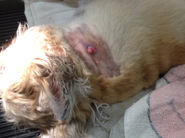
The lump on Derby's neck.

Charlie, the veiled chameleon.

The growth bubble on Charlies head (the green and brown striped sphere is his eye, and the brown-grey sphere right above that is the growth bubble).
Pete Pebbles was the only surgery
(besides a declaw) that we had today, and it was a major surgery. He was brought in today for vomiting, and
while he was being checked on the doctors realized he had a broken leg. According to his owner, he had been limping
for about five weeks, so his leg had probably broken five weeks ago. However, the owner claimed to not even know
of such a circumstance, which makes no sense to me; and it irritates me that the
owner realized something was wrong but chose to do nothing about it, because
now the animal has been in unnecessary pain for this long of a time. The leg was fractured at the knee, and since
this happened five weeks ago the bone naturally started to heal on its
own. Also naturally (and unfortunately),
the bone healed very crookedly, and in the wrong area. Dr. Mark took the cat into surgery to fix the
leg. He gently ran his blade through the
skin, tissue, and muscle to the bone, and then spent a bit trying to cut and
pull off the scar tissue so he could directly access the bone. Once he got to the bone and contemplated
what he was dealing with, he re-broke the knee so he could fix it
properly. However, because the bone had started
to heal so crookedly, the edges of bone that had been rubbing together were
extremely crooked now and he couldn’t get them to stay together because they
kept slipping and moving; therefore, he placed a pin in the kneecap to keep the
bone together so it would heal properly.
Stephanie got him a sterilized drill and pin, and Dr. Mark diligently worked
at getting the pin in over the course of about 45 minutes (he considered
putting two pins in, but the bone is fairly small and he didn’t want to take
the chance of damaging the bone, or having the pins conflict with each other). The pin was quite long compared to the size
of the cat. He drilled the bone all the
way through the thigh bone (and through the skin up at the top of his thigh;
the pin has to go all the way through the leg to ensure that the pin is at the
correct angle. If it’s even slightly
off, it could ruin both the bone and the procedure), and he worked on regaining
control of the pin before switching sides and starting to slightly drill back
the way he came so the pin would also go into the bones at the knee (he called
this a fracture grade [going through the thigh bone], reduce [gaining control],
and normal grade back [gently and partially going back the way he came from to
connect the two bones with the pin]).
After he connected the two bones with the pin, he moved the leg to make
sure everything looked okay worked properly, which everything did. Dr. Mark then cut off the excess pin sticking
out of Pete Pebbles’ leg, and sewed up the surgical incision and the hole
caused by the pin going through his leg.
After bandaging and wrapping the leg, we placed him in recovery on some
blankets so he would be fairly comfortable and relaxed when he woke up.

Dr. Mark slicing through the skin, tissue, and muscle of Pete Pebbles' knee to access the bone.

Cutting and pulling away all the scar tissue on the bone.

A view of all the different muscle and tissue layers. The light purple/grey triangle in the middle is the bone.

Another view of the different layers and bone.

After re-breaking the bone; the bone here is sticking up, by Dr. Mark's thumb.

Getting ready to drill the pin into the bone.

Drilling the pin into the bone.

Regaining control of the pin before/while switching sides.

The pin all the way through the thigh bone (which is again sticking out with the left side of the pin). As I had said, it was a long pin compared to the cat.


Stitching the incision up.

Cutting off the excess pin before stitching that small hole up (Dr. Mark had to make the hole that the pin made in the skin a little bigger because the skin was getting caught/wrapped in the drill, so to stop that he made the hole a little bigger, as will be seen in the next picture).

The finished stitching.

Fitting a plastic full-leg splint.

Starting to wrap the leg.

Finishing the under dressing of the bandaging.

The finalized wrapping with stiff Vet wrap covering the under bandaging.
No comments:
Post a Comment